Vascular/Doppler Ultrasound

let us helping you by make an appointment

Lorem ipsum dolor sit amet consectetur.

What is Vascular/Doppler Ultrasound?

Vascular ultrasound provides pictures of the body’s veins and arteries. Doppler ultrasound is a special ultrasound technique that evaluates blood flow through a blood vessel, including the body’s major arteries and veins in the abdomen, arms, legs and neck. Doppler ultrasound measures the direction and speed of blood cells as they move through vessels. The movement of blood cells causes a change in pitch of the reflected sound waves (called the Doppler effect). A computer collects and processes the sounds and creates graphs or color pictures that represent the flow of blood through the blood vessels.
What are some common uses for Vascular/Doppler exams:

Vascular ultrasound is performed to:
  • Help monitor the blood flow to organs and tissues throughout the body.
  • Locate and identify blockages (stenosis) and abnormalities like plaque or emboli.
  • Detect blood clots (deep venous thrombosis (DVT)) in the legs or arms.
  • Determine whether a patient is a good candidate for a procedure like angioplasty.
  • Determine if there is an enlarged artery (aneurysm).
  • Determine the source and severity of varicose veins.
Doppler ultrasound images can help the physician evaluate:
  • Blood flow blockages (such as clots).
  • Narrowing vessels.
  • Tumors and congenital malformation.

How is the procedure performed?

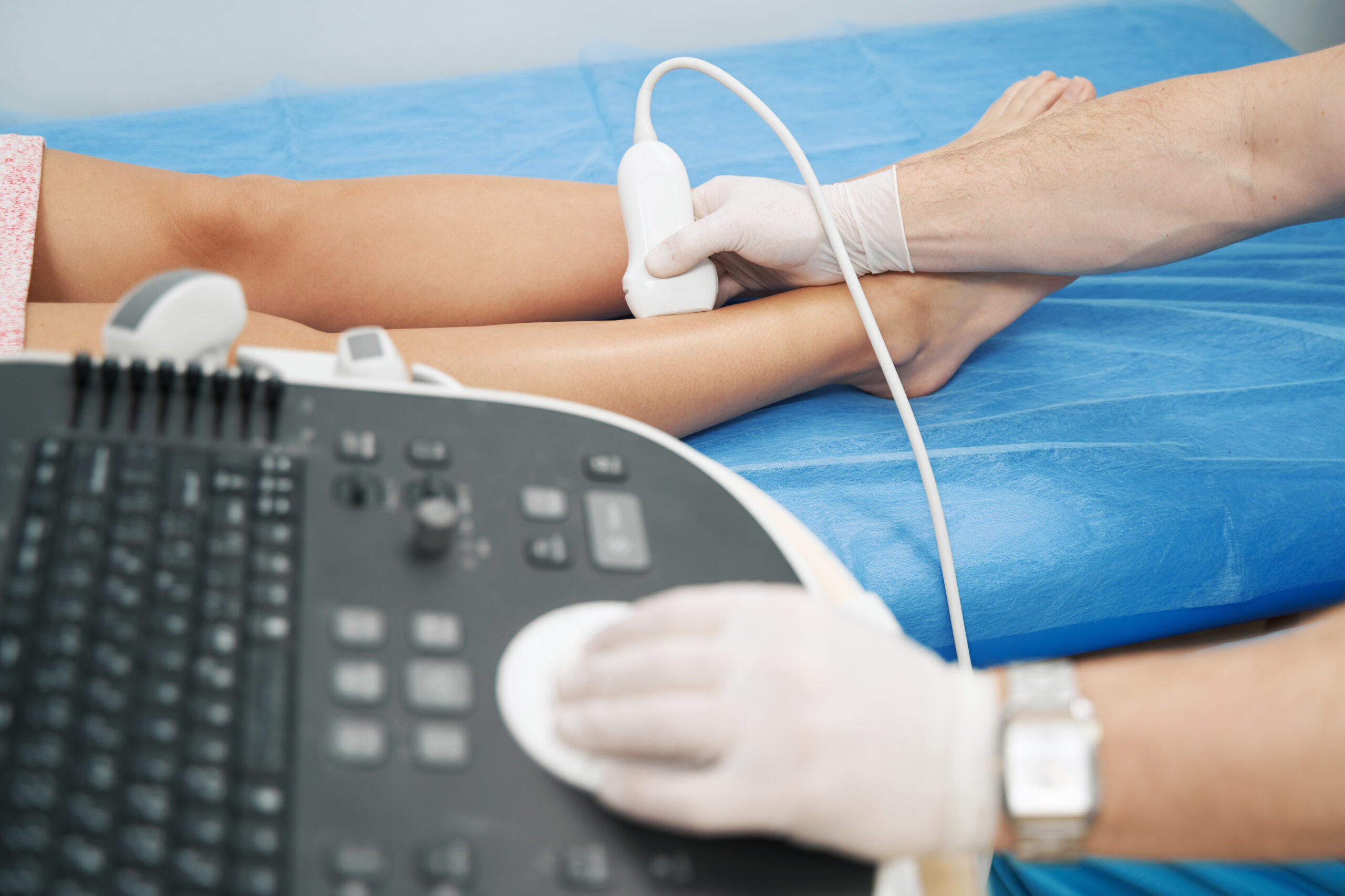
Just like General Ultrasound Vascular/Doppler examinations are painless, fast and easy. After you are positioned on the examination table, sonographer will apply some warm gel on your skin and then place the transducer against your body, moving it back and forth over the area of interest until the desired images are captured. There is usually no discomfort from pressure as the transducer is pressed against the area being examined. Once the imaging is complete, the gel will be wiped off your skin.

Who interprets the results and how do I get them?

A Board Certified Radiologist trained to interpret radiology exams will analyze the images and send them to our PACS system. From our PACS system the ordering physician and nurse will be able to view the images and the report from any computer using or HIPPA compliant log in screen. No one, but your ordering physician and your nurse, will be able to see your report.
work hours & numbers